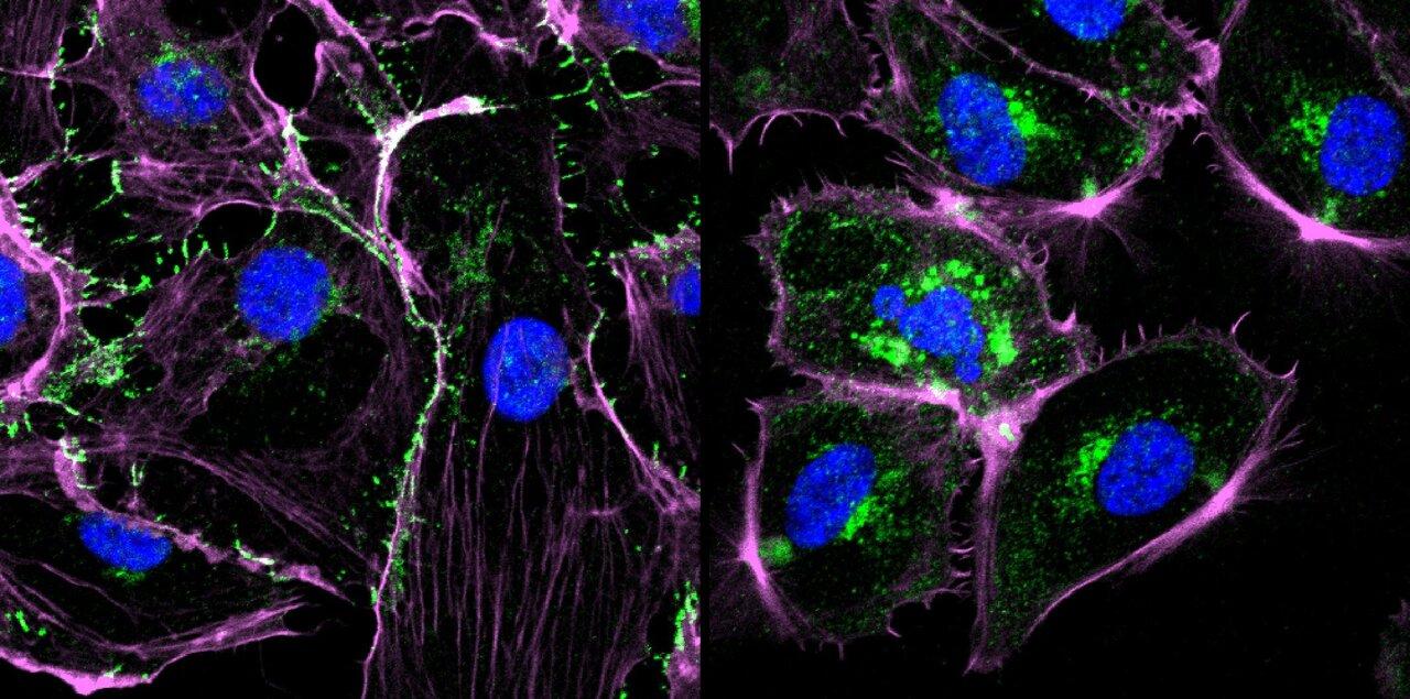
Cell migration and invasion play a crucial role in many physiological and pathological processes. During embryonic development, migration of cells is essential for proper formation of tissues and organs. Post-embryonically, migration of immune cells is important for immune surveillance and response. However, uncontrolled migration and invasion of cells also contribute to disease progression in cancers, atherosclerosis, and rheumatoid arthritis. In vitro assays help researchers study and understand the complex mechanisms underlying cell migration and invasion in normal and disease conditions. They also enable screening of potential anti-metastatic drugs.
Traditional Cell Migration And Cell Invasion Assay
One of the simplest ways to study Cell Migration And Cell Invasion Assay is the wound healing or scratch assay. In this assay, a gap or wound is created in a monolayer of adherent cells using a pipette tip or similar device. Microscopic images are then captured at regular intervals to track the movement of cells and closure of the wounded area. While easy to perform, this assay does not mimic cell migration in 3D physiologically relevant environments.
Another traditional method used is the Boyden chamber or transwell assay where cells are seeded on a porous membrane insert placed inside a well. A chemoattractant or chemorepellent is added in the lower chamber to create a gradient. Cells that migrate through the pores toward or away from the chemoattractant/repellent over a period of time are fixed, stained and counted under a microscope. Though more physiologically relevant than scratch assays, transwell assays only assess two-dimensional migration of cells.
Modern Cell Migration Assays
To better model cell migration in more complex 3D environments like the extracellular matrix (ECM), advances have been made in developing extracellular matrix-based assays. In Ibidi μ-slide chemotaxis chambers, cells embedded in ECM are monitored microscopically in the presence of defined chemical gradients. Another approach involves seeding cells inside 3D ECM protein gels like collagen or fibronectin and monitoring their migration using time-lapse microscopy over hours to days.
Microfluidic devices have also enabled exquisite control over chemotactic environmental cues and real-time high-resolution imaging of single cell migration dynamics in 3D spaces with ECM components. For example, the collagen-microchannels platform guides cell migration along defined lines within a 3D collagen matrix. These modern assays provide quantitative data on various migration parameters like speed, persistence, turning angles along with guidance cues. They can assess migration of cancer cells, stem cells, immune cells or fibroblasts in native-like environments.
Cell Invasion Assays
While cell migration refers to movement of single cells on extracellular surfaces, cell invasion involves active penetration and passage of cells through ECM and basement membrane barriers. In vitro invasion assays primarily use reconstituted basement membrane extract models like Matrigel to study this process.
In the traditional Matrigel invasion assay, Matrigel is layered over a porous membrane in a transwell insert. Cells are seeded on top and allowed to actively invade through the Matrigel layer toward a chemoattractant in the lower chamber over 24-72 hours. Non-invading cells on the upper surface are removed and invaded cells on the lower surface are stained, imaged and counted.
Although physiologically relevant, traditional Matrigel invasion assays are laborious, low-throughput and quantification is challenging. To overcome these limitations, 3D spheroid invasion models and microfluidic-based assays have been developed recently. In the 3D spheroid model, multicellular spheroids generated from cancer or other cell types are embedded in 3D matrices like Matrigel. Time-lapse imaging tracks the expansion and penetration of invading cells from the spheroid core over time. Microfluidic devices have complex 3D matrix-filled channels that enable real-time monitoring of live cell invasion under tunable gradients. Such advanced platforms provide quantitative data on 3D cell motility and invasion parameters.
Applications And Significance
Cell migration and invasion assays are invaluable research tools that aid in understanding normal development and disease progression at the cellular and molecular levels. They play a crucial role in cancer metastasis research by facilitating identification of key genes, pathways and microenvironmental factors regulating cancer cell dissemination. Such assays enable screening of potential anti-metastatic or anti-invasive drugs.
They are also used to study cell migration mechanisms in other diseases like atherosclerosis, rheumatoid arthritis as well as in regenerative processes during wound healing. By closely mimicking the physiological 3D in vivo microenvironment, advanced modern assays provide more accurate and predictive insights compared to traditional methods. Overall, cell migration and invasion assays continue to improve our understanding of cell behaviours and help translate basic science discoveries into effective clinical applications and therapies.
Get more insights on this topic: https://savagesoulhub33.blogspot.com/2024/09/cell-migration-and-cell-invasion-assay_4.html
Author Bio
Vaagisha brings over three years of expertise as a content editor in the market research domain. Originally a creative writer, she discovered her passion for editing, combining her flair for writing with a meticulous eye for detail. Her ability to craft and refine compelling content makes her an invaluable asset in delivering polished and engaging write-ups. (LinkedIn: https://www.linkedin.com/in/vaagisha-singh-8080b91)
*Note:
1. Source: Coherent Market Insights, Public sources, Desk research
2. We have leveraged AI tools to mine information and compile it

