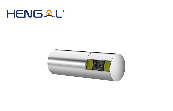
What is the working principle of the industrial endoscope camera module? The imaging principle of the electronic industrial endoscope camera module is to use the light emitted by the light source equipped in the TV information center to guide the light into the cavity of the subject through the light guide fiber in the endoscope, and the CCD image sensor It receives the light reflected from the mucosal surface of the body cavity, converts the light into electrical signals, and then transmits the signals to the TV information center through wires, and then stores and processes these electrical signals through the TV information center, and then transmits them to the TV monitor. A color mucosal image of the inspected cavity is displayed on the screen. There are two types of CCD image sensors currently used in the world, and their specific ways of forming color images are slightly different.
industrial endoscope camera module
First of all, what aspects should I start with when configuring a set of imaging diagnostic workstations (CR, DR, CT, MRI) or configuring a PACS, RIS system medical display? How to choose an industrial endoscope camera module reasonably? Let's learn together!
industrial endoscope camera module
1. Select from the function
The optional industrial endoscope camera module that can perform DICOM correction has special correction software; there is an optical sensor interface on the back of the display, which can be connected to the optical sensor for correction, otherwise the correction cannot be performed. The medical display with constant brightness device is selected to ensure that the brightness of the display does not change with time, and it can ensure the consistency and integrity of the system display. Due to the requirements of teaching and the habits of doctors, doctors at home and abroad are accustomed to use pens to point on the film or display screen to express their views on the specific details of the image. The LCD screen material is a fragile material. In order to adapt to the medical environment, display manufacturers will Responsibly install the protective plate of the LCD screen at the time of production. Therefore, the optional medical display should have a protective plate for the LCD screen.
2. Select from the parameters
The diagnostic workstation is recommended to be equipped with 3MP and 5MP monitors, and 3MP is the main choice for those without mammography and flat-panel DR. Observation and teaching workstations are recommended to be equipped with 2MP and 1MP monitors. The optional medical monitor has a dedicated graphics card with 10bit output grayscale.
3. Choose from certification
Optional medical certification: FDA510(k), ISO13485 certification, safety certification: CE, UL, CCC certification is considered to be on the industrial endoscope camera module. CCC certification is China's mandatory safety certification, if only CCC certification should not be used in the medical field.
What is the influence of the electronic industrial endoscope camera module on the imaging? The electronic endoscope hole probe image is often affected by noise and appears not clear enough. There are many reasons for the noise, such as the internal noise of sensitive components, the particle noise of photosensitive materials, and thermal noise. , jitter noise caused by relative motion, interference noise of transmission channel, quantization noise, etc. Reflected on the image, the noise makes the originally uniform and continuously changing gray scale suddenly increase or decrease, forming some isolated points and false edges, and sometimes even submerging the features, which brings a lot of difficulties to the analysis. The cause of noise determines the distribution characteristics of noise and its relationship with the image signal.
The DSP digital processing circuit reduces signal distortion and ensures that the electronic industrial endoscope camera module can get high-quality images. Automatic white balance and excellent color adjustment function ensure the true reproduction of electronic endoscope images. The contour is enhanced, and the metering range can be adjusted as needed. The automatic electronic shutter ensures well-exposed, well-defined images in any situation. What are the components of the industrial endoscope camera module?
What are the components of the industrial endoscope camera module? Endoscopes can be divided into two types: rigid endoscopes and flexible endoscopes, also known as rigid endoscopes and flexible endoscopes. Rigid endoscope includes three parts: image transmission, illumination and air hole. The image transmission part is divided into objective lens, relay system and eyepiece to form a transmission image. The lighting part adopts the method that the cold light source uses the optical fiber to penetrate into the interior. The function of the stoma is for air supply, water supply and biopsy forceps. Rigid endoscope products include hysteroscope, proctoscope, hysteroscope, thoracoscope and so on.
How to choose the industrial endoscope camera module reasonably?
The industrial endoscope camera module that uses fiber beam imaging and light guide or uses CCD to guide the image becomes a flexible endoscope. It has been widely used in medicine because of its good softness and convenient operation performance. The current products include gastroscope, duodenoscope, colonoscope, choledochoscope, enteroscope, bronchoscope, nasopharyngoscope, ureteroscope, etc.
The characteristics of the flexible endoscope are: it is soft and can easily enter the complex internal organs of the human body, which not only reduces the pain of the patient, but also reaches places that cannot be reached by the rigid endoscope. With the head bending mechanism, the blind spot can be eliminated. Sampling and treatment can be done through the biopsy hole.
The soft industrial endoscope camera module can be divided into fiber endoscope and electronic endoscope. How to hold the flexible endoscope: wear gloves, hold it with both hands, hold the grip part in one hand and the lens body in the other hand, do not let the apex sag, and hold it firmly. Fiber endoscope structure: tip part, bending part, insertion part, operation part, light guide hose, light guide connection part, eyepiece. The apex is formed into a small rigid section, with direct-view (front-view), side-view, and strabismus. The gastroscope and colonoscopy adopt the direct view method, and the duodenoesophagoscope adopts the side view method. The top of the front end is provided with: objective lens hole (image guide beam), light hole (light guide beam), air-water hole (nozzle), and biopsy hole.
industrial endoscope camera module https://www.hengal-tech.com/3-7mm-industrial-endoscope-camera-module-3-7mm-pshow/15.html

