Introduction
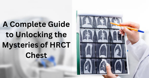
In the realm of medical diagnostics, High-Resolution Computed Tomography (HRCT) chest scans stand as a vital tool in the assessment of various pulmonary conditions. From identifying lung disorders to evaluating treatment efficacy, HRCT chest scans provide clinicians with detailed insights into the thoracic region. This comprehensive guide aims to demystify HRCT chest scans, offering a thorough exploration of the procedure, its significance, and interpretation of findings.
Understanding HRCT Chest Scans
HRCT chest scans, short for High-Resolution Computed Tomography of the chest, are advanced imaging procedures used to examine the structures within the thoracic cavity in great detail. These scans offer a comprehensive view of the chest area, allowing healthcare professionals to detect and diagnose various pulmonary conditions with precision.
What is HRCT Chest?
HRCT chest, which stands for High-Resolution Computed Tomography of the chest, is a specialized medical imaging procedure used to obtain detailed images of the structures within the thoracic cavity. Unlike traditional X-rays, HRCT chest scans provide high-resolution cross-sectional images of the chest area, allowing healthcare professionals to visualize the lungs, heart, and surrounding tissues with exceptional clarity and precision.
These scans utilize advanced technology to generate multiple cross-sectional images of the chest, which are then reconstructed into three-dimensional representations of the internal anatomy. By capturing images from various angles, HRCT chest scans enable radiologists to detect and evaluate a wide range of pulmonary conditions, including lung nodules, interstitial lung diseases, pulmonary embolism, and lung cancer.
HRCT chest scans are particularly valuable for assessing the presence of abnormalities such as nodules, ground-glass opacities, consolidations, and fibrotic changes within the lungs. These findings help clinicians in diagnosing respiratory conditions, monitoring disease progression, and planning appropriate treatment strategies for patients.
Significance of HRCT Chest
HRCT chest scans hold immense significance in the field of diagnostic imaging, particularly in the assessment of pulmonary conditions. These specialized scans offer detailed insights into the chest anatomy, allowing healthcare professionals to detect and diagnose a wide range of respiratory disorders with precision. The significance of HRCT chest scans stems from several key factors:
Detailed Visualization: HRCT chest scans provide high-resolution images of the chest area, including the lungs, heart, and surrounding structures. This detailed visualization enables radiologists to identify subtle abnormalities and assess the extent of disease involvement accurately.
Diagnostic Accuracy: By capturing cross-sectional images of the chest, HRCT scans allow for the detection of various pulmonary abnormalities, such as lung nodules, ground-glass opacities, and fibrotic changes. This diagnostic accuracy is essential for distinguishing between different lung conditions and guiding appropriate treatment decisions.
Early Detection: HRCT chest scans are instrumental in the early detection of lung cancer, interstitial lung diseases, and other pulmonary conditions. Early diagnosis facilitates prompt initiation of treatment, leading to better outcomes and improved patient survival rates.
Assessment of Disease Progression: In patients with chronic respiratory conditions, such as pulmonary fibrosis or chronic obstructive pulmonary disease (COPD), HRCT chest scans are invaluable for monitoring disease progression over time. These scans provide longitudinal data on changes in lung morphology, helping clinicians assess treatment response and adjust therapeutic interventions as needed.
Preoperative Evaluation: Before thoracic surgeries or lung transplant procedures, HRCT chest scans play a crucial role in preoperative planning and assessment. These scans provide detailed anatomical information, allowing surgeons to evaluate pulmonary anatomy, identify potential surgical risks, and optimize surgical approaches for improved outcomes.
Research and Education: HRCT chest scans are indispensable tools for research and educational purposes in the field of respiratory medicine. These scans contribute to the advancement of medical knowledge by facilitating studies on disease pathophysiology, treatment efficacy, and imaging techniques.
The procedure of HRCT Chest
The procedure for performing an HRCT (High-Resolution Computed Tomography) chest scan involves several steps to ensure accurate and detailed imaging of the thoracic cavity. Here's a breakdown of the typical procedure:
Patient Preparation: Before the scan, patients may be instructed to change into a hospital gown and remove any metallic objects, such as jewelry, watches, or clothing with metal fasteners. It's essential to inform the healthcare provider about any pre-existing medical conditions, allergies, or medications.
Positioning: The patient lies flat on a motorized table that moves through the center of the CT scanner. It's crucial to maintain a still position during the scan to minimize motion artifacts and ensure clear imaging.
Breath-Holding Instructions: To obtain high-quality images, patients may be asked to hold their breath for a few seconds during specific parts of the scan. This helps reduce respiratory motion and improves the clarity of the images.
Scanning Process: Once the patient is positioned correctly, the CT scanner rotates around the body, emitting X-ray beams from multiple angles. These beams pass through the chest and are detected by sensors on the opposite side of the scanner.
Image Acquisition: As the scanner rotates, it captures multiple cross-sectional images (slices) of the chest at various depths. These images are acquired in thin sections, typically ranging from 1 to 5 millimeters, to ensure high-resolution imaging.
Contrast Administration (if applicable): In some cases, a contrast agent may be administered intravenously to enhance the visibility of certain structures or abnormalities within the chest. This contrast material may be iodine-based and is usually injected through a vein in the arm.
Post-Processing: After the scan is complete, the raw image data is processed by a computer to reconstruct detailed cross-sectional images of the chest. These images are then reviewed and interpreted by a radiologist or other healthcare professional trained in diagnostic imaging.
Patient Discharge: Once the scan is finished, patients can usually resume their normal activities immediately. It's essential to follow any post-scan instructions provided by the healthcare provider, such as drinking plenty of fluids to flush out any contrast material.
Preparation for HRCT Chest
Patients undergoing HRCT chest scans are typically advised to refrain from eating for a few hours before the procedure. It is crucial to inform the healthcare provider about any existing medical conditions or allergies.
Interpretation of HRCT Chest Findings
Interpreting the findings of an HRCT (High-Resolution Computed Tomography) chest scan requires expertise and attention to detail. Radiologists analyze the images carefully to identify and assess various pulmonary abnormalities, providing valuable diagnostic insights. Here's an overview of the key aspects involved in the interpretation of HRCT chest findings:
Density and Texture: Radiologists evaluate the density and texture of lung parenchyma on HRCT images. Normal lung tissue appears dark (low density) on the scan, while abnormalities may manifest as areas of increased or decreased density. Ground-glass opacities, consolidations, and nodules are common findings that warrant further evaluation.
Lung Nodules: HRCT chest scans are sensitive in detecting lung nodules, which are small, round, or oval-shaped lesions within the lung parenchyma. Radiologists assess the size, shape, and characteristics of nodules to determine their likelihood of malignancy. Features such as spiculation, irregular margins, and increased density may indicate a higher risk of cancer.
Ground-Glass Opacities (GGOs): GGOs appear as hazy or cloudy areas of increased lung density on HRCT scans. These findings may indicate a wide range of pulmonary conditions, including infections, inflammation, or early-stage lung cancer. Radiologists assess the distribution, extent, and characteristics of GGOs to guide further diagnostic evaluation and treatment planning.
Consolidations: Lung consolidations represent areas of lung tissue that have become solidified due to various pathological processes, such as pneumonia, atelectasis, or pulmonary edema. On HRCT scans, consolidations appear as regions of increased density with air bronchograms or vascular markings. Radiologists analyze the distribution, shape, and associated findings to determine the underlying cause of consolidation.
Fibrotic Changes: HRCT chest scans are valuable for evaluating fibrotic changes within the lungs, such as pulmonary fibrosis or interstitial lung diseases (ILDs). Fibrotic changes appear as linear or reticular opacities on the scan, indicating scarring and remodeling of lung tissue. Radiologists assess the pattern, distribution, and extent of fibrosis to differentiate between different ILDs and monitor disease progression.
Pleural Abnormalities: HRCT scans also allow for the assessment of pleural abnormalities, such as pleural effusions, pleural thickening, or pleural plaques. These findings may indicate underlying lung pathology or pleural disease, requiring further investigation and management.
Clinical Applications of HRCT Chest
Diagnosis of Interstitial Lung Diseases
HRCT chest plays a crucial role in diagnosing interstitial lung diseases (ILDs), facilitating the identification of patterns indicative of specific ILDs such as idiopathic pulmonary fibrosis (IPF) or sarcoidosis.
Screening for Lung Cancer
Screening for lung cancer is a crucial aspect of preventive healthcare, especially for individuals at high risk of developing this deadly disease. Lung cancer screening aims to detect lung nodules or tumors at an early stage when treatment options are most effective, potentially improving patient outcomes and reducing mortality rates.
Conclusion
In conclusion, HRCT chest scans serve as invaluable tools in the diagnosis and management of various thoracic conditions. By providing detailed imaging of pulmonary structures, HRCT chest scans empower healthcare professionals to make informed decisions regarding patient care and treatment strategies. Understanding the nuances of HRCT chest scans enables patients and clinicians to navigate the diagnostic process confidently, fostering better health outcomes.
FAQs
What is the difference between HRCT chest and conventional CT scans?
- HRCT chest offers higher spatial resolution, making it ideal for evaluating pulmonary structures in greater detail compared to conventional CT scans.
Is HRCT chest a painful procedure?
- No, HRCT chest scans are non-invasive and painless. Patients may experience mild discomfort lying still during the scan.
Are there any risks associated with HRCT chest scans?
- While HRCT chest scans involve exposure to ionizing radiation, the benefits of accurate diagnosis outweigh the potential risks, especially in the context of medical necessity.
How long does an HRCT chest scan take?
- An HRCT chest scan typically ranges from 10 to 30 minutes, depending on the complexity of the examination.
Can HRCT chest scans detect lung cancer?
- Yes, HRCT chest scans are highly sensitive in detecting lung nodules, aiding in the early detection and diagnosis of lung cancer.
Are there any alternatives to HRCT chest scans?
- In certain cases, alternative imaging modalities such as MRI or ultrasound may be considered based on the clinical indication and patient factors.

